


導入事例 Odyssey M イメージングシステム

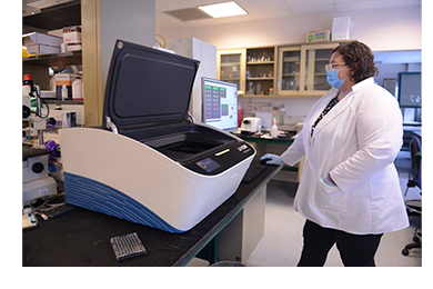
Dr. Kimberley Samkoe
Develops Quantitative Fluorescence Imaging Methodologies for Applications in Oncology
Having long been fascinated by the interaction of light with molecules and tissues, Dr. Kimberley Samkoe uses fluorescence imaging to provide microscopic molecular information that can be interpreted at a macro-scale for applications in oncology. As Principal Investigator at the Samkoe Lab in the Center for Imaging Medicine in the Thayer School for Engineering at Dartmouth College, she is motivated to create highly sensitive, specific, and quantitative fluorescence imaging methodologies that aim to streamline clinical procedures such as surgical resection and therapeutic decision-making.
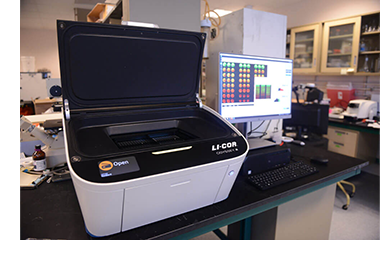
Dr. Samkoe and her lab have used the Odyssey CLx Imaging System extensively in their research for over a decade, and now use the Odyssey M for imaging freshly excised tissue sections, fixed-cell based assays, pathology slide imaging, In-Cell Western assays, and Western blots.
❝ The Odyssey M is a joy to use. Not only is it user-friendly for both new and experienced researchers, but the wide array of options available for both fluorescence and RGB Truecolor imaging enables a flexibility in data collection that no other system we know of provides. ❞
- Dr. Kimberley Samkoe
Fluorescent Imaging in Cancer Research
The Samkoe Lab’s research goal is to develop quantitative clinical methodologies to improve clinical decision-making around surgical resection and molecular-targeted therapy. These include fluorescence imaging methodologies that will streamline surgical resection of cancerous tissue by utilizing fluorescent Affibody molecules, which are rapidly taken up by tissue of interest and cleared by normal tissue. This results in suitable contrast within hours, allowing imaging and surgical resection to take place on the same day.
Currently, their research focuses on developing fluorescence guided surgery applications of single-agent and paired-agent methodologies in pre-clinical and clinical applications, as well as studying receptor occupancy and therapeutic response of cancers to a variety of molecular therapeutic agents.
The lab uses the Odyssey M in their research for in vitro live cell assays to assess fluorescent molecular targets, and In-Cell Western™ assays to validate live cell molecular imaging and to detect changes within cellular signaling pathways. Additionally, they extensively image fluorescence molecular targets in fresh, fresh-frozen, and fixed tissues at a macroscopic scale.
❝ The time savings of having all these features in a single instrument, that negates extensive post-image processing required when using different instruments, is tremendous. ❞
- Dr. Kimberley Samkoe
The Odyssey M Helps Overcome Challenges

Dr. Kimberley Samkoe
One of the biggest challenges her lab has faced is in the alignment of fluorescent images with RGB Truecolor images of thinly sectioned (4-7 mm) fresh frozen, formalin fixed, and pathologically stained tissues. The limitations of instruments unable to image visible and near-infrared fluorescence in addition to white-light or RGB Truecolor imaging in the same orientation and resolution underly this challenge.
The ability of the Odyssey M to image in many fluorescent and RGB Truecolor combinations improves their imaging procedures and helps meet this challenge. Samkoe’s lab can collect both fluorescent and RGB Truecolor images on fresh tissue and on pathological slides using both the epi- and trans-illumination functions. The capability of the Odyssey M to image at a range of resolutions—5, 10, 20, 50 and 100 µm—increases their ability to detect residual cancer in both fixed and fresh tissue.
A Transformation in Data Collection
The Odyssey M streamlines the Samkoe Lab’s ability to collect, align, and analyze data from fresh, fresh-frozen, and fixed tissues. The ability to collect both fluorescence and RGB Truecolor images from the same, or consecutive sample(s) using identical resolution and imaging parameters has transformed the way they collect data and has greatly reduced the time it takes to do so.
Dr. Samkoe finds that Empiria Studio Software—developed for use with Odyssey Imaging Systems—provides an excellent base for experimental planning, execution, and analysis, especially for cell-based assays and In-Cell Westerns, where plate template creation aids in experimental planning and laboratory teaching. Reproducibility of data day-to-day, ease of use, and ease of documentation give Dr. Samkoe confidence in her data.
We thank Dr. Samkoe for her significant contributions to cancer research and are proud to call her an Odyssey expert.
Photo credit: Kathryn LoConte Lapierre, Associate Director of Creative Services, Editor, Dartmouth Engineer Magazine
資料ダウンロード
- Wang, C., Xu, X., Folaron, M., Gunn, J. R., Hodge, S., Chen, E. Y., Hoopes, P. J., Tichauer, K. M., & Samkoe, K. S. (2021, October 14). Improved discrimination of tumors with low and heterogeneous EGFR expression in fluorescence-guided surgery through paired-agent protocols. Molecular Imaging and Biology. https://doi.org/10.1007/s11307-021-01656-3.
- Wang, C., Xu, X., Hodge, S., Chen, E. Y., Hoopes, P. J., Tichauer, K. M., & Samkoe, K. S. (2021, October 12). Identification of a suitable untargeted agent for the clinical translation of aby-029 paired-agent imaging in fluorescence-guided surgery. Molecular Imaging and Biology. https://doi.org/10.1007/s11307-021-01642-9.
- Souza, A. L. R. de, Marra, K., Gunn, J., Samkoe, K. S., Hull, S., Paulsen, K. D., & Pogue, B. W. (2018, September 28). Optimizing glioma detection using an egfr‐targeted fluorescent affibody. Photochemistry and Photobiology. https://doi.org/10.1111/php.13003.
- Samkoe, K. S., Bates, B. D., Elliott, J. T., LaRochelle, E., Gunn, J. R., Marra, K., Feldwisch, J., Ramkumar, D. B., Bauer, D. F., Paulsen, K. D., Pogue, B. W., & Henderson, E. R. (2018). Application of fluorescence-guided surgery to subsurface cancers requiring wide local excision. Cancer Control, 25(1), 107327481775233. https://doi.org/10.1177/1073274817752332.
- Elliott, J. T., Marra, K., Evans, L. T., Davis, S. C., Samkoe, K. S., Feldwisch, J., Paulsen, K. D., Roberts, D. W., & Pogue, B. W. (2017, May 1). Simultaneous in vivo fluorescent markers for perfusion, protoporphyrin metabolism, and EGFR expression for optically guided identification of orthotopic glioma. Clinical Cancer Research. https://doi.org/10.1158/1078-0432.CCR-16-1400.
- Samkoe, K. S., Tichauer, K. M., Gunn, J. R., Wells, W. A., Hasan, T., & Pogue, B. W. (2014, December 15). Quantitative in vivo immunohistochemistry of epidermal growth factor receptor using a receptor concentration imaging approach. Cancer Research. https://doi.org/10.1158/0008-5472.CAN-14-0141.
- Kanick, S. C., Tichauer, K. M., Gunn, J., Samkoe, K. S., & Pogue, B. W. (2014, August 29). Pixel-based absorption correction for dual-tracer fluorescence imaging of receptor binding potential. Biomedical Optics Express. https://doi.org/10.1364/BOE.5.003280.
- Sexton, K., Tichauer, K., Samkoe, K. S., Gunn, J., Hoopes, P. J., & Pogue, B. W. (2013, April 12). Fluorescent affibody peptide penetration in glioma margin is superior to full antibody. PLOS ONE. https://doi.org/10.1371/journal.pone.0060390.
製品/シリーズに関するお問い合わせ
製品に関するご質問や技術的なお問い合わせなどはこちらのフォームをご利用ください。